
PET Scan, as one of the most advanced medical imaging techniques, enables the detection of cellular and metabolic changes, assisting physicians in diagnosing cancer with greater accuracy, earlier, and in a more targeted manner. This imaging method not only identifies the presence of tumors but also provides valuable information about the activity and spread of cancer cells to the treatment team.
What is a PET scan?
PET scan, or Positron Emission Tomography, is an advanced imaging technique that, instead of merely displaying the structure of organs, examines the function and metabolism of cells. In this method, a small amount of radioactive material (usually a labeled sugar) is injected into the body. Cells with higher activity, such as cancer cells, absorb more of this substance and appear as active regions in the PET scan images.
How does a PET scan help in diagnosing cancer?
Cancer cells have a higher energy consumption compared to healthy cells. PET scans, by detecting this increased consumption, can:
- To distinguish active tumors from healthy tissues.
- Better distinguishes between benign and malignant lesions.
- Even very small cellular changes can be detected before they become visible on a CT scan or MRI.
For this reason, PET scans are considered one of the most accurate tools for diagnosing and monitoring cancer.
Applications of PET scans in cancers
PET scans are utilized at various stages of cancer-related diseases, including:
Early diagnosis of cancer
In cases where other imaging methods fail to provide definitive results, PET scans can assist in identifying suspicious lesions.
Determining the Stage of Cancer (Staging)
A PET scan determines the extent to which cancer has spread in the body and whether the lymph nodes or other organs are involved.
Evaluation of response to treatment
After chemotherapy or radiotherapy, a PET scan determines whether cancer cells are still active or if the treatment has been effective.
Diagnosis of cancer recurrence
In patients with a history of cancer, PET scans can detect the recurrence of the disease at its early stages.
The difference between PET scan, CT scan, and MRI.
While CT scans and MRIs primarily display the anatomical structure of the body, PET scans focus on cellular activity.
| Feature | PET scan | CT scan | MRI |
| Type of Examination | Cellular Function | Body structure | Soft tissue |
| Precision in Cancer Diagnosis | Very high | Moderate | Average |
| Tumor Activity Diagnosis | has | Does not exist. | Limited |
In many advanced centers, this method is performed as PET/CT to simultaneously evaluate both functional and structural information.
How is a PET scan performed?
Undergoing a PET scan (Positron Emission Tomography) is a non-invasive, precise, and step-by-step process that is typically painless and highly safe. This imaging technique is specifically designed to clearly examine the activity of the body’s cells, particularly cancerous cells.The steps for performing a PET scan are as follows:
1. Injection of radioactive material
Initially, a very small amount of a labeled radioactive substance (usually sugar) is introduced into the body through intravenous injection.
This substance is harmless and specifically accumulates in cells with higher metabolic activity.
2. Waiting period for substance absorption
After the injection, the patient should remain at rest for approximately 30 to 60 minutes to allow the injected substance to distribute properly within the body.
During this time, physical movement or activity is not recommended, as it may affect the accuracy of the images.
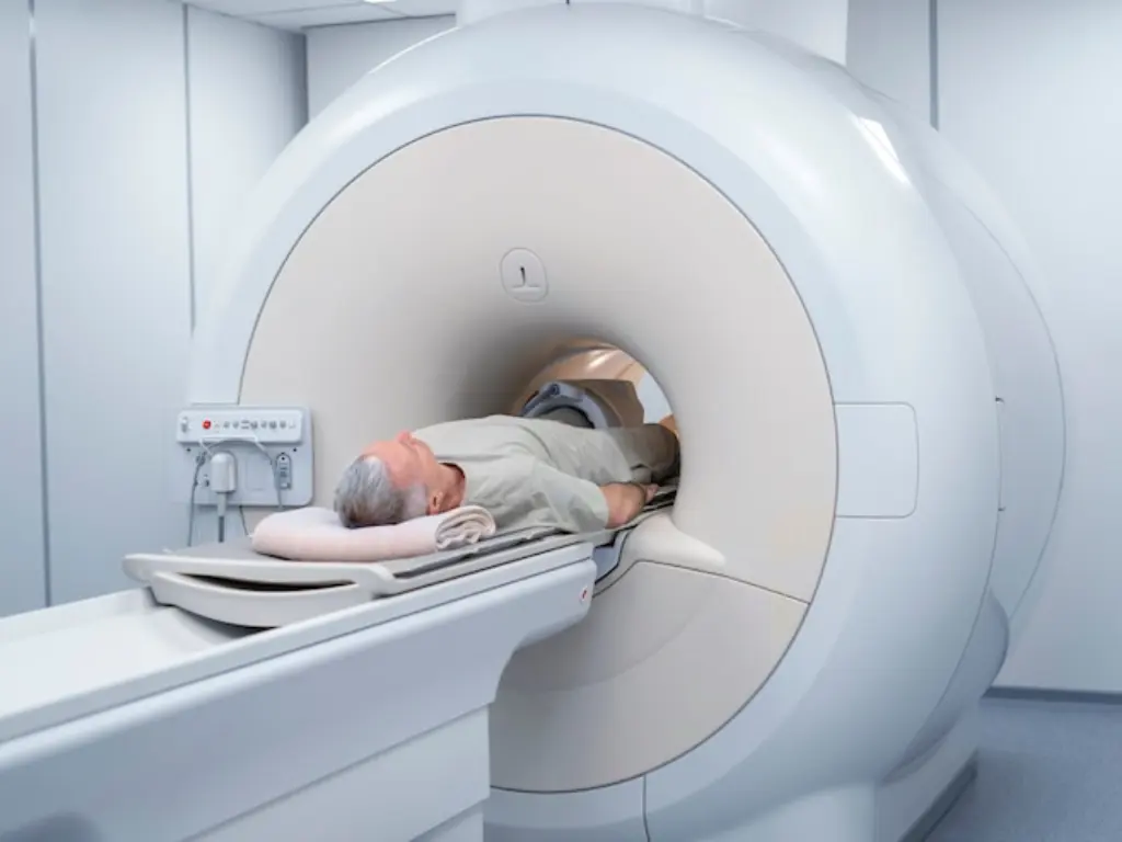
3. Perform imaging
The patient lies down on the specialized PET scan bed, and the bed slowly moves into the machine. At this stage:
- Imaging is completely painless.
- The patient must remain still for a few minutes.
- The device detects areas of the body with higher cellular activity.
- The duration of imaging is usually between 20 to 45 minutes.
4. Completion of Work and Interpretation of Images
After the imaging procedure is completed, the patient can resume their daily activities. The obtained images are reviewed and interpreted by a specialized radiologist, and the final results are provided to the treating physician. Following the PET scan, it is recommended to drink plenty of fluids to help expedite the elimination of the injected substance from the body.
Necessary Preparations Before Undergoing a PET Scan
Observing the preparation guidelines before undergoing a PET Scan plays a crucial role in ensuring the accuracy of imaging results. Neglecting these instructions can lead to reduced image quality or incorrect interpretation. The most important preparation points include:
Fasting
The patient must fast for at least 6 hours prior to undergoing the PET scan. During this time, consuming food and sugary drinks is prohibited; however, drinking plain water is allowed.
Avoid physical activity
Avoid engaging in intense exercise or physical activity for at least 24 hours prior to your PET scan, as muscle activity can alter the absorption of the injected substance.
Blood sugar control
In patients with diabetes, precise blood sugar control is highly important. Elevated blood sugar levels may reduce the accuracy of PET scans. Any adjustment to medication or insulin must be made strictly under the supervision of a physician.
Medication usage
In most cases, taking regular medications is not an issue. However, it is essential to provide the list of medications you are using to the doctor or the center’s staff prior to undergoing a PET scan.
Information about pregnancy or breastfeeding
If you are pregnant or breastfeeding, it is essential to inform your doctor prior to undergoing a PET scan so that appropriate decisions can be made.
Personal attire and belongings
- It is recommended to wear comfortable clothing without any metal components.
- Before imaging, metallic objects such as jewelry, watches, and mobile phones must be removed.
On the day of the PET scan, it is best for the patient to remain completely calm and follow the imaging center’s instructions to ensure the highest quality images are obtained.

Is a PET scan dangerous?
The amount of radiation used in a PET scan is controlled and safe. The radioactive substance is eliminated from the body within a short period, and when medically necessary, the benefits of this method far outweigh its potential risks.
Advantages of PET Scan in Cancer Diagnosis
PET scan (Positron Emission Tomography) is one of the most precise imaging methods for diagnosing and evaluating cancer. Unlike many other techniques, it does not rely solely on the physical appearance of tissues but examines cellular activity and tumor metabolism. The key advantages of PET scans in cancer diagnosis include:
Early Detection of Cancer
A PET scan can detect metabolic changes in cells at the early stages of a disease, even when a mass is not yet visible on a CT scan or MRI.
Precise differentiation between benign and malignant lesions
By evaluating cellular activity, PET scans assist physicians in making a more precise distinction between benign and malignant lesions, thereby preventing unnecessary examinations.
Precise determination of the cancer stage (Stage)
PET scans play a crucial role in determining the extent of cancer spread within the body and can accurately detect involvement of lymph nodes or distant organs.
Evaluation of response to treatment
After chemotherapy or radiotherapy, a PET scan determines whether cancer cells are still active or if the treatment has been effective, even before any visible changes in tumor size occur.
Diagnosis of cancer recurrence
In patients with a history of cancer, PET scans can detect the recurrence of the disease at early stages, enabling a more rapid initiation of treatment.
Reducing unnecessary diagnostic procedures
By improving diagnostic accuracy, PET scans can reduce the need for repetitive or unnecessary biopsies and imaging procedures.
PET scans, by providing detailed information about the activity of cancer cells, are considered an invaluable tool for early diagnosis, accurate staging, and effective monitoring of cancer treatment. They play a crucial role in guiding treatment decisions.
Which patients are candidates for undergoing a PET scan?
A PET scan (Positron Emission Tomography) is not typically prescribed for everyone but is utilized in specific medical situations where the information obtained can play a significant role in diagnosis or treatment decision-making. The primary groups considered candidates for a PET scan include:
Patients with cancer
PET scans are one of the primary tools for evaluating various types of cancer. They are used for initial diagnosis, determining the stage of the disease, and monitoring the progress of treatment in these patients.
Patients with a suspicious mass or lesion
In cases where a mass is detected in the body but its nature cannot be definitively determined through other methods, PET scans can assist in identifying whether the lesion is benign or malignant.
Patients who require evaluation for the spread of cancer.
To assess lymph node involvement or the spread of cancer to other organs, a PET scan provides the physician with precise information.
Patients undergoing or post cancer treatment
PET scans are used to evaluate the effectiveness of chemotherapy, radiotherapy, or targeted therapies, and to determine whether cancer cells are still active.
Patients at risk of cancer recurrence
In individuals with a history of cancer who exhibit suspicious symptoms or findings, PET scans can detect disease recurrence at its early stages.
Some cardiac or cerebral patients (in specific cases)
In certain cases, a PET scan is also prescribed to assess the viability of heart muscle or to evaluate specific neurological disorders.
The final decision to perform a PET scan is always made by the treating physician based on the patient’s clinical condition, test results, and findings from other imaging methods.
Limitations and Contraindications of PET Scan
Despite the high accuracy of PET scans in diagnosing diseases, this imaging method has certain limitations in specific situations and may not be suitable for everyone. Being aware of these factors helps ensure informed and safe decision-making.
Pregnancy
During pregnancy, PET scans are generally not recommended due to the use of radioactive material, except in specific and absolutely necessary situations where the physician determines that the benefits outweigh the potential risks.
Breastfeeding period
In breastfeeding mothers, PET scans are performed with caution, and it is generally recommended to temporarily stop breastfeeding for a specific period after the imaging procedure. The exact duration of this interruption is determined by the physician.
Uncontrolled blood sugar
In diabetic patients with high and uncontrolled blood sugar levels, the accuracy of PET scan results decreases because the uptake of the injected substance is affected. Under such conditions, performing the PET scan may need to be postponed.
Inability to remain still
Patients who are unable to remain still during imaging due to physical issues, severe pain, or anxiety may face limitations in undergoing a PET scan.
Certain specific medical conditions
In rare cases, certain metabolic disorders or the use of specific medications can affect the accuracy of the results, necessitating further evaluation before performing a PET scan.
PET scans are a safe and accurate method, but it is essential to evaluate the patient’s individual condition before performing the procedure. Consulting with the treating physician and providing detailed information about the patient’s health status are the best ways to make an informed decision regarding undergoing a PET scan.

Frequently Asked Questions About PET Scans
Does a PET scan cause pain?
No, PET scan is a non-invasive and painless procedure. The only part that might cause slight discomfort is the injection of the radioactive substance at the beginning, which is similar to a simple intravenous injection. The imaging process itself is completely painless, and the patient only needs to lie still on the device’s table for a few minutes.
How long does a PET scan take?
The entire PET scan process typically takes between 1.5 to 2 hours. This duration includes the injection of the tracer, a waiting period of approximately 30 to 60 minutes for the substance to be absorbed by the body, and the imaging procedure itself. The imaging portion alone usually takes less than an hour.
Is PET scan radiation harmful to the body?
The amount of radiation used in a PET scan is controlled and safe, adhering to medical standards. The radioactive substance used is eliminated from the body within a short period. If prescribed by a physician, the diagnostic benefits of a PET scan far outweigh its potential risks.
What care is necessary after undergoing a PET scan?
After undergoing a PET scan, there are usually no specific restrictions, and the patient can resume their daily activities. It is recommended to drink plenty of fluids to help flush the injected substance from the body more quickly. Additionally, following the specific instructions provided by the imaging center is important.
Is the result of a PET scan alone sufficient for diagnosis?
A PET scan provides highly detailed information about cellular activity; however, the final interpretation is always performed by a specialist physician in conjunction with other tests and imaging studies. In some cases, a PET scan serves as a complementary diagnostic tool, aiding in more accurate decision-making.
Summary
PET scan is one of the most precise and advanced medical imaging techniques, playing a crucial role in the diagnosis, staging, and monitoring of cancer treatment. Conducting this examination in well-equipped centers under the supervision of specialized physicians can clarify the treatment pathway and enhance the chances of successful outcomes.